Foto
Video
Audio
Vektoren
Benutzername:
Passwort:
Passwort vergessen
Kostenlose Registrierung
Foto
Video
Audio
Vektoren
Mein Profil
Warenkorb
(
0
)
Leuchtkästen
( 0 )
schnelle Hilfe
en
de
en
us
eu
fr
es
mx
Erweiterte Suche
Suchbegriff
Urheber
Suchbegriff ausschliessen
Abo Filter
Nur ClipDealer
Social-Media Lizenz
Mediengattung
Video
Audio
Photo
Vector
Verwendungszweck
Kommerzielle Verwendung
Redaktionelle Verwendung
Menschen
Personen ausschließen
1 Person
2 Personen
3 Personen
ab 4 Personen
Farbe
Farbe
Schwarzweiß
Sepia
Farbwähler
#
Visuell ähnlich
Visuell ähnlich
Bildformat
Hochformat
Querformat
Einstellung
Makro
Licht
Morgen
Tag
Abend
Nacht
Position
Augenhöhe
Aufsicht
Untersicht
Außenaufnahme
Innenaufnahme
Luftaufnahme
Unterwasseraufnahme
Auflösung
mindestens L, 4 MP
mindestens XL, 8 MP
mindestens XXL, 15 MP
mindestens XXXL, 30 MP
Datum
Alle
≤ 2 Jahre
≤ 1 Jahr
≤ 6 Monate
≤ 1 Monat
≤ 1 Woche
Kategorie
Abstrakt
Architektur
Brunnen
Brücken
Denkmäler
Details
Dächer
Fenster
Tore & Türen
Fachwerk
Hochhäuser
Häuser & Gebäude
Industrieanlagen
Innenarchitektur
Zimmer & Räume
Kirchen & religiöse Bauten
Mühlen
Park- & Gartenanlagen
Plätze
Ruinen
Schlösser & Burgen
Städte & Dörfer
Türme
Berufe & Branchen
Businesskonzepte
Dienstleistungen
Film & Fernsehen
Handel & Verkauf
Handwerk & Bau
Industrie
Informatik
Landwirtschaft
Logistik & Transport
Medizin & Pharma & Labor
Polizei & Recht
Presse
Schule & Bildung
Verwaltung
Wissenschaft & Forschung
Essen & Trinken
Fisch & Meeresfrüchte
Fleisch & Wurst
Früchte & Obst
Gemüse & Salat
Gerichte
Getränke
alkoholische Getränke
Kaffee
Tee
Gewürze & Kräuter
Kuchen & Gebäck
Snacks & Fast Food
Süßigkeiten & Knabbereien
Fahrzeuge & Verkehr
Flugverkehr
Schienenverkehr
Schifffahrt
Straßenverkehr
Verkehrszeichen
Finanzen & Börse
Aktien & Wertpapiere
Geldscheine & Münzen
Freestyle
Buchstaben & Zahlen
Comic & Illustrationen
Zeichen & Symbole
Freizeit
Events
Erntedank
Geburtstag
Halloween
Karneval
Ostern
Silvester
Valentinstag
Weihnachten
Gastronomie
Kino
Konzerte
Kunst
Nachtleben
Religion
Spiele
Theater & Bühne
Wohnen
Gesundheit & Beauty
Diät
Körperpflege & Kosmetik
Medikamente
Medizinische Geräte
Wellness
Hintergründe & Effekte
Länder & Regionen
Afrika
Asien
Australien
Europa
Deutschland
England
Finnland
Frankreich
Griechenland
Italien
Norwegen
Portugal
Schweden
Schweiz
Spanien
Österreich
Mittelamerika / Karibik
Nordamerika
Südamerika
Menschen
Akt
Baby & Schwangerschaft
Familien
Frauen
Gruppen
Kinder & Jugendliche
Körperteile
Männer
Pärchen
Senioren
Natur & Umwelt
Berge & Gebirge
Blätter & Blumen
Bäume & Wälder
Felder & Wiesen
Flüsse & Seen
Himmel & Wolken
Luftaufnahmen
Meer & Strand
Mineralien & Steine
Naturkatastrophen
Sonnenaufgang & -untergang
Unterwasser
Weltall & Astronomie
Wind & Wetter
Wüsten
Ökologie
Objekte & Details
Deko & Stillleben
Fahnen & Flaggen
Kleidung
Musikinstrumente
Möbel
Schmuck & Uhren
Sport
Ballsport
Bergsport
Extremsport
Fitness & Kraftsport
Gymnastik & Turnen
Kampfsport
Laufen & Walken
Leichtathletik
Luftsport
Motorsport
Radsport
Reitsport
Wassersport
Wintersport
Technik
Audio
Computer
Elektronik
Energie & Strom
Maschinen & Werkzeuge
Telekommunikation
TV & Video
Tiere
Amphibien
Fische & Krustentiere
Insekten & Spinnentiere
Mollusken
Reptilien
Säugetiere
Haus- und Nutztiere
Meeres- und Süßwassersäuge
Primaten
Wildtiere
Vögel
Es ist kein Fehler aufgetreten
Hinweis!
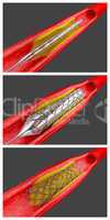
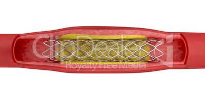
164 Bilder zum Thema "myocardial ischemia" bei ClipDealer
alle Filter zurücksetzen
Suchbegriff:
myocardial
Suchbegriff:
ischemia
Suchbegriff:
myocardial ischemia
Medien sortieren nach
Datum
Bewertungen
Aufrufen
Relevanz
Download
aufsteigend
absteigend
Gehe zu Seite:
« Vorherige
1
2
3
Nächste »
Nächste Seite »
« Vorherige
1
2
3
Nächste »