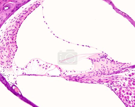
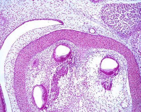
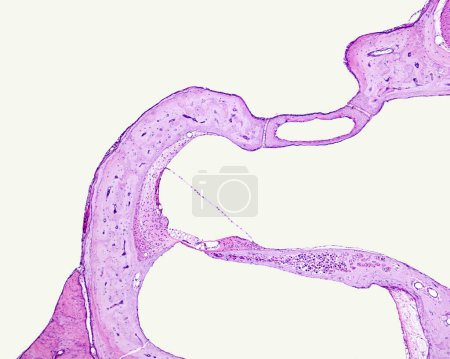
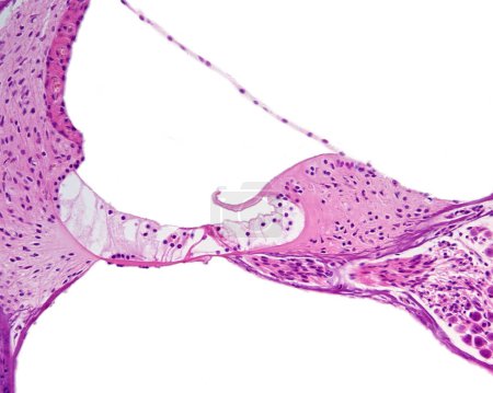
4 Bilder zum Thema "cochlear duct" bei ClipDealer

Medien sortieren nach     Gehe zu Seite:    
« Vorherige  1  Nächste »
Light micrograph of a cross section of the cochlea of the inner ear showing from top to bottom: vestibular, cochlear and tympanic ducts or scala , separated by Reissner membrane and basilar membrane. The cochlear duct shows, from right to left, the s
Light microscope micrograph showing three sections of a developing cochlea where can be already recognized the epithelial thickening that will be the organ of Corti and the future spiral ganglion. The bony labyrinth of temporal bone is still in carti
Low magnification light microscope micrograph showing, on the left, the organ of Corti covered by the Reissner membrane. On top, the oval window occluded by the base of the stapes can be seen
Light micrograph of a cross section of the cochlea scala media or cochlear duct showing from right to left: limbus spiralis with tectorial membrane and Reissner membrane, organ of Corti with tunnel of Corti and hair cells, and the stria vascularis.

« Vorherige  1  Nächste »