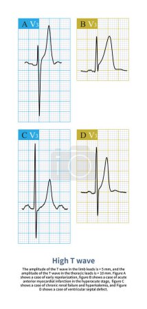
44 Bilder zum Thema "Repolarisation" bei ClipDealer

Medien sortieren nach     Gehe zu Seite:    
« Vorherige  1  Nächste »
When the amplitude of the T wave exceeds 5 mm in the limb leads and 10 mm in the thoracic leads, it is called high T wave.High T waves can be seen in physiological and pathological conditions.
3D illustration of an electrocardiogram (ECG) showing prolonged QT interval with broad-based T-waves, characteristic of type 1 long QT syndrome.
Long QT syndrome, type 2. 3D illustration of an electrocardiogram (ECG) showing prolonged QT interval with notched and of lower amplitude T-waves.
At present, there is a younger trend in patients with acute myocardial infarction, so it is important to check the ECG for acute chest pain in young people.
3D illustration of an electrocardiogram (ECG) showing prolonged QT interval with broad-based T-waves, characteristic of type 1 long QT syndrome.
Early repolarization is a common benign ECG change that manifests as J-point ECG with or without ST-segment elevation.
3D illustration of an electrocardiogram (ECG) showing prolonged QT interval with broad-based T-waves, characteristic of type 1 long QT syndrome.
Hypoplastic left heart syndrome is a rare congenital heart defect in which the left side of the heart is severely underdeveloped.It may affect the left ventricle, aorta, aortic valve, or mitral valve. 3D Render
3D illustration of an electrocardiogram (ECG) showing prolonged QT interval with broad-based T-waves, characteristic of type 1 long QT syndrome.
Long QT syndrome, type 2. 3D illustration of an electrocardiogram (ECG) showing prolonged QT interval with notched and of lower amplitude T-waves.
The presence of ST-segment prolongation and T wave symmetry and high tip on ECG suggests hypocalcemia and hyperkalemia.
3D illustration of an electrocardiogram (ECG) showing prolonged QT interval with broad-based T-waves, characteristic of type 1 long QT syndrome.
Long QT syndrome, type 2. 3D illustration of an electrocardiogram (ECG) showing prolonged QT interval with notched and of lower amplitude T-waves.
Hypoplastic left heart syndrome is a rare congenital heart defect in which the left side of the heart is severely underdeveloped.It may affect the left ventricle, aorta, aortic valve, or mitral valve. 3D Render
3D illustration of an electrocardiogram (ECG) showing prolonged QT interval with broad-based T-waves, characteristic of type 1 long QT syndrome.
The atrial muscle and atrioventricular node belong to fast and slow response cells, respectively, and phase 0 depolarization is responsible for Na+ channels and Ca2+ channels, respectively.
The autonomic  frequency of the sinoatrial node is the fastest, and other secondary pacemakers are controlled through mechanisms of preemptive occupation and overspeed suppression.
3D illustration of an electrocardiogram (ECG) showing prolonged QT interval with broad-based T-waves, characteristic of type 1 long QT syndrome.
Long QT syndrome, type 2. 3D illustration of an electrocardiogram (ECG) showing prolonged QT interval with notched and of lower amplitude T-waves.
Firstly, select point J as the reference point, and then select 60ms after point J as the measurement point to evaluate the ST segment offset morphology and amplitude.
Long QT syndrome, type 2. 3D illustration of an electrocardiogram (ECG) showing prolonged QT interval with notched and of lower amplitude T-waves.
3D illustration of an electrocardiogram (ECG) showing prolonged QT interval with broad-based T-waves, characteristic of type 1 long QT syndrome.
Long QT syndrome, type 2. 3D illustration of an electrocardiogram (ECG) showing prolonged QT interval with notched and of lower amplitude T-waves.
3D illustration of an electrocardiogram (ECG) showing prolonged QT interval with broad-based T-waves, characteristic of type 1 long QT syndrome.
Normal T waves are asymmetrical. Coronary T waves are not absolutely symmetric, but increase in symmetry. From the bottom of the T wave to the bottom, the asymmetry gradually increases.
3D illustration of an electrocardiogram (ECG) showing prolonged QT interval with broad-based T-waves, characteristic of type 1 long QT syndrome.
Long QT syndrome, type 2. 3D illustration of an electrocardiogram (ECG) showing prolonged QT interval with notched and of lower amplitude T-waves.
The QT interval of ECG is from the beginning of QRS wave to the end of T wave, representing the total time of ventricular depolarization and repolarization.
R wave greater than S wave is judged to be positive; R smaller than S  is judged to be negative; R equal to S amplitude is judged to be equipotential.
3D illustration of an electrocardiogram (ECG) showing prolonged QT interval with broad-based T-waves, characteristic of type 1 long QT syndrome.

« Vorherige  1  Nächste »