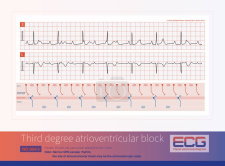
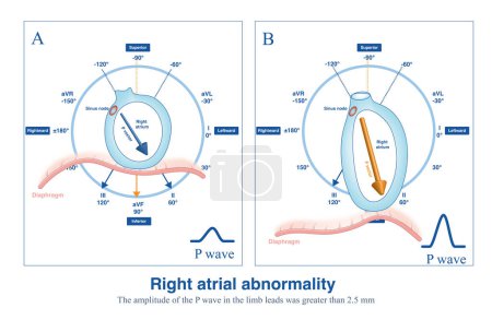
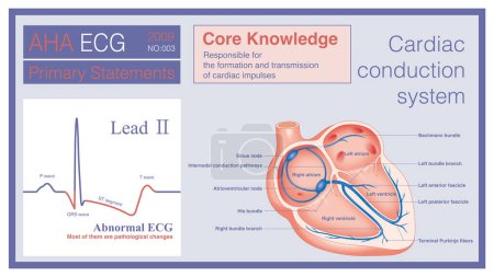
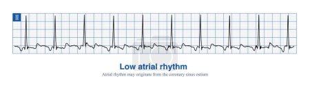
10316062 Bilder zum Thema "ECG monitoring" bei ClipDealer

Medien sortieren nach     Gehe zu Seite:    
« Vorherige  1  2  3  4  5  6  7  ...  171935  
Monitoring Icon
Diagnosis against blue medical background with dna and ecg
Monitoring Icon
Monitoring Icon
Monitoring Icon
Monitoring Icon
Monitoring Icon
Monitoring Icon
Monitoring Icon
Pulse Monitoring Icon
Composite image of diabetic woman using blood glucose monitor
ECG
Pulse Monitoring Icon
Cardio Tablet Means Online Www And Wellness
ECG
ECG
ECG
ECG
ECG
ECG
ECG
ECG
ECG
ECG
Closeup of electrocardiogram EKG printout
Cardio Workouts Shows Getting Fit And Beat
heart with life line
Heartbeat Tablet Means Pulse Trace And Cardiology
Health Heartbeat Shows Beating Well And Jog 3d Rendering
Cardio Fitness Indicates Physical Activity And Cardiogram
Health Heartbeat Represents Wellness Sprint And Render 3d Render
Heartbeat Online Means Pulse Trace And Cardiac
Heart Monitor
digital pixel EKG electrocardiogram blue background
heart with cardiograph
Closeup of electrocardiogram printout being held
healthy heart
Heartbeat Monitor
happy hearts
healthy heart
heart beats
wounded heart with bandage
heart with cardiology
Flatline Heart Monitor - Alert
In acute high lateral myocardial infarction, there is indicative ST segment elevation in leads I and aVL, and corresponding ST segment depression in leads II, III and aVF.
Heart and EKG ECG Graph
ECG electrodes on the patient
Medical doctor making ECG test in hospital.
The illustration shows the two patterns of ventricular tachycardia episodes.The green circle represents sinus rhythm. Picture A shows paroxysmal episodes of ventricular tachycardia, and picture B shows short bursts.
Bidirectional ventricular tachycardia is a kind of malignant arrhythmia. The polarity of QRS main wave alternates from beat to beat, and it is easy to degenerate into ventricular fibrillation.
A 36 year old man survived CPR after sudden syncope. The electrocardiogram was suggestive of Brugada syndrome type 1. Implantation of ICD therapy.
R wave greater than S wave is judged to be positive; R smaller than S  is judged to be negative; R equal to S amplitude is judged to be equipotential.
Sometimes, because the QRS axis is in the upper left quadrant, the high-amplitude R wave of left ventricular hypertrophy occurs in the limb leads, and left chest leads is normal.
ECG monitoring of patient
The QT interval of ECG is from the beginning of QRS wave to the end of T wave, representing the total time of ventricular depolarization and repolarization.
During the onset of variant angina pectoris, ECG is divided into non fusion wave, partial fusion wave and complete fusion wave according to the fusion degree of QRS wave, ST segment and T wave.
Ecg monitor recording activity of heart rate during hospital operation, surgery
Female, 51 years old, diagnosed with mitral stenosis. When this ECG was taken, the patient still maintained sinus rhythm.Note that the P wave duration was widened.
In case of acute anterior myocardial infarction, the characteristics of ST segment elevation in ECG can be used to deduce whether the culprit vessel system is the left main trunk or the proximal LAD.
Medical diagnostics in modern hospital.
The left main coronary artery can be divided into the left anterior descending artery and the left circumflex artery, and sometimes the intermediate branch artery.
The standard for diagnosing right atrial abnormality in ECG is that the amplitude of P-wave in limb leadsI is greater than 2.5mm, and the amplitude of upright P-wave in chest leads is  1.5mm.
Third degree atrioventricular block in young women may be congenital, with the block located on the atrioventricular node or above bifurcation of the His bundle.
When sinus arrest occurs, the electrocardiogram will show a long P-P interval, which is not multiples of the basal sinus cycle, including physiological and pathological reasons.
When the rhythm of the atria originates in the lower part of the atria, the whole atria are excited from inferior to superior, producing negative P waves in the inferior leads.
Male, 84 years old, admitted to hospital with chest pain for 1 day. These ECG rhythms are the Holter monitor records of the patients after admission, and they are third degree atrioventricular block.
Four anatomical malformations of tetralogy of Fallot: 1 aortic straddling; 2 ventricular septal defect; 3 right ventricular hypertrophy and 4 pulmonary artery stenosis.
Third degree atrioventricular block in young women may be congenital, with the block located on the atrioventricular node or above bifurcation of the His bundle.
Male, 71 years old, was clinically diagnosed with upper gastrointestinal bleeding. During sleep at night, ECG monitoring showed sinus bradycardia, blood pressure 115 and 70mmHg.
In patients with emphysema, the anatomical position of the heart is more vertical, the depolarization potential of the right atrium from top to bottom increases, and the P wave of the ECG increases.
electronic sensor on chest for Holter monitoring, track pacemaker dysfunction, daily monitoring of electrocardiogram, blood pressure, cardiac examination, treatment of cardiac diseases
sensors on chest for Holter monitoring, woman with Holter monitor daily monitoring of electrocardiogram, blood pressure, cardiac examination, treatment of cardiac diseases, track pacemaker dysfunction
Abnormal ECG refers to changes in depolarization waves and or repolarization waves, most of which are pathologic and few are physiological.
When the rhythm of the atria originates in the lower part of the atria, the whole atria are excited from inferior to superior, producing negative P waves in the inferior leads.
When ectopic impulses from the anterior wall of the right atrium produce a completely negative P wave in lead V1, the posterior wall ectopic impulse produces a positive and negative biphasic P wave.
When ectopic focal areas in the atria are located in the atrial septum, the left atrium and right atrium can be excited at the same time, producing a very narrow P wave.
When the ECG is too complex, the recording quality is poor, or it is impossible to interpret what is causing the loss of the ECG, the ECG machine will not be able to provide a diagnostic reference.
According to the offset amplitude of the ST segment at J point and J60 point, ST segment elevation can be divided into three basic types: concave upward, oblique straight and concave downward type.
Male, 13 years old, clinically diagnosed with secundum atrial septal defect. Note that the QRS wave in lead V1 of the electrocardiogram has a qR shape, indicating right ventricular hypertrophy.
Third degree atrioventricular block in young women may be congenital, with the block located on the atrioventricular node or above bifurcation of the His bundle.
Sometimes, there may be slight non-specific changes and normal variations in the electrocardiogram, which are often due to physiological reasons and have no clinical therapeutic significance.
Abnormal ECG refers to changes in depolarization waves and or repolarization waves, most of which are pathologic and few are physiological.
When the duration of the P wave exceeds 120 ms, the amplitude of the limb leads exceeds 2.5 mm, and the amplitude of the thoracic leads exceeds 1.5 mm, it is interpreted as a biatrial abnormality.
When intraatrial excitation is transmitted from one atrium to the other, a wider duration of P waves are generated, such as sinus rhythm, lateral  left atrial focal, etc.
A 4-year-old boy with a clinical diagnosis of long QT syndrome. No genetic testing was done during hospitalization. The child died suddenly during follow-up.
Sometimes, it is necessary to place electrodes in the high intercostal space at the conventional chest lead electrode placement position, which is called a high intercostal chest lead.
Firstly, select point J as the reference point, and then select 60ms after point J as the measurement point to evaluate the ST segment offset morphology and amplitude.
Female, 5-year-old, clinically diagnosed with dextrocardia. The characteristic of dextrocardia electrocardiogram is a gradual decrease in R-wave amplitude from leads V1 to V6.
When intraatrial excitation is transmitted from one atrium to the other, a wider duration of P waves are generated, such as sinus rhythm, lateral  left atrial focal, etc.
Abnormal ECG refers to changes in depolarization waves and or repolarization waves, most of which are pathologic and few are physiological.
When the ECG is too complex, the recording quality is poor, or it is impossible to interpret what is causing the loss of the ECG, the ECG machine will not be able to provide a diagnostic reference.
sensors on female body for Holter monitoring, woman with Holter monitor for daily monitoring of electrocardiogram, blood pressure, cardiac examination, treatment of cardiac diseases
Male, 10 years old, was clinically diagnosed with tetralogy of Fallot. ECG shows an elevated P wave amplitude in thoracic leads, suggesting right atrial abnormality.
A 67-year-old man presents with heart palpitations, numbness of the lips and nausea after consuming poisonous shellfish. ECG showed sinus bradycardia.

« Vorherige  1  2  3  4  5  6  7  ...  171935